In our lab we analyze cells and tissue using a computational imaging technique known as Fourier ptychographic microscopy. We have built a Fourier ptychography microscope in our lab to quantitatively image the optical phase delay of biological samples. Quantitative phase imaging is a recent development in optical microscopy that allows the sample's intrinsic physical properties to be measured. The quantitative phase imaging technique involves imaging the phase delay of the light wave at each location of the sample, generating a 2 dimensional map. This phase delay map can be interpreted as a mass density map of the sample, a new observable we wish to correlate with disease. Traditionally dye staining is used to generate contrast in cell and tissue samples for imaging. The staining procedure and subsequent color imaging is prone to laboratory dependent variations however, causing issues in reproducibility and automated analysis. The quantitative phase imaging approach is attractive from two points of view: 1. it bypasses the color dependent variabilities of stained sample color imaging, 2. it provides a new avenue of investigation of robust optical phase based biomarkers based on the cell/tissue sample's intrinsic physical properties.
The Fourier ptychography microscope we are using for this purpose is similar to a regular light microscope except that the illumination source is replaced by a programmable LED array. The LED array allows images of the sample to be captured using illumination from different angles. These images are then computationally combined to create a high-resolution quantitative phase image. We aim to find quantitative phase based biomarkers that can be used to diagnose and stage pathological cells and tissue. We work in collaboration with the Pathology department of Bezmialem Vakif University Hospital.
We are also investigating the application of deep learning and machine learning algorithms to automate and improve disease diagnosis and staging in digital pathology. Images may be used to train neural networks that can find correlations that go unnoticed by even expert practitioners. By harnessing these correlations in image classification we wish to enhance the diagnostic power of medical caregivers, thereby helping to improve the quality of healthcare.
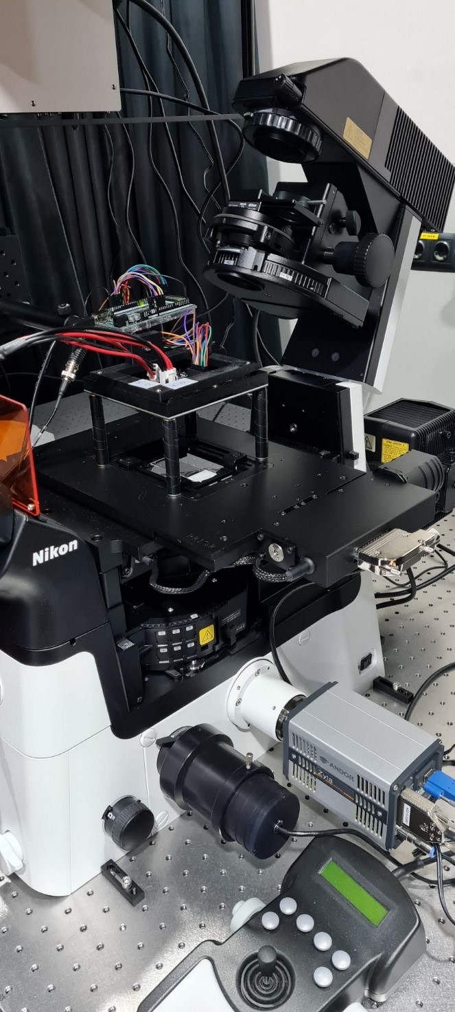
Fourier ptychography microscope for quantitative phase imaging at BILSAB.
Projects:
- Liver fibrosis staging using quantitative phase imaging and machine learning.
Teaching:
- Biophysics
Graduate Student Supervision:
- MSc, Lutfi Kadir Çelebi, Istanbul Technical University, 2021-present. Liver fibrosis staging using quantitative phase imaging and machine learning. (co-advisor)
Lab Research Team:
- Lutfi Kadir Çelebi Technician
Contact Info:
E-mail: oakcakir@bezmialem.edu.tr